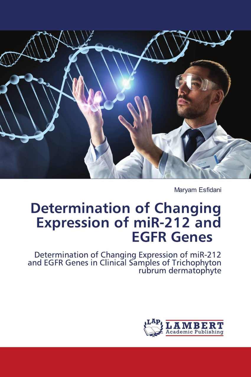
کتاب Determination of Changing Expression of miR-212 and EGFR Genes
۳۷۸,۰۰۰ تومان قیمت اصلی: ۳۷۸,۰۰۰ تومان بود.۱۶۳,۸۰۰ تومانقیمت فعلی: ۱۶۳,۸۰۰ تومان.
| تعداد صفحات | 85 |
|---|---|
| شابک | 978-620-3-02933-8 |
| انتشارات |

کتاب Determination of Changing Expression of miR-212 and EGFR Genes – کشف نقش تغییرات ژنی در بیماریها
کتاب Determination of Changing Expression of miR-212 and EGFR Genes اثری علمی و پژوهشی است که به بررسی تغییرات بیان ژنهای miR-212 و EGFR و نقش آنها در بیماریهای مختلف میپردازد. این کتاب بهطور جامع تأثیرات این تغییرات را بر فرایندهای سلولی، از جمله رشد، تمایز و پاسخ به درمان، تحلیل میکند.
درباره کتاب Determination of Changing Expression of miR-212 and EGFR Genes
این اثر پژوهشی به بررسی نقش miR-212 و EGFR به عنوان عوامل کلیدی در تنظیم مکانیسمهای مولکولی در بیماریها، بهویژه سرطان، اختصاص دارد. نویسنده با تحلیل دادههای آزمایشگاهی و مطالعات بالینی، اطلاعات جامعی درباره تعامل این دو عامل ژنی و تأثیرات آنها بر رشد سلولها و پاسخ به درمان ارائه میدهد.
موضوعات کلیدی کتاب
- نقش miR-212 در بیماریها: بررسی اثرات تنظیمی این microRNA بر مسیرهای زیستی و مولکولی.
- EGFR و عملکرد آن: تحلیل نقش گیرنده فاکتور رشد اپیدرمال در فرایندهای سلولی و پیشرفت بیماریها.
- تعامل miR-212 و EGFR: مطالعه ارتباط متقابل این دو عامل و تأثیر آنها بر درمانهای هدفمند.
- روشهای نوین تحقیقاتی: معرفی تکنیکهای پیشرفته برای مطالعه بیان ژنی.
ویژگیهای برجسته کتاب Determination of Changing Expression of miR-212 and EGFR Genes
- تحقیقات دقیق و علمی: ارائه نتایج پژوهشهای جدید و معتبر در زمینه زیستشناسی مولکولی.
- بینش کاربردی: بررسی نقش تغییرات ژنی در توسعه درمانهای نوین و بهبود راهکارهای بالینی.
- ساختار منسجم: کتاب بهگونهای سازماندهی شده که خوانندگان بهراحتی بتوانند مفاهیم کلیدی را درک کنند.
- تأکید بر جنبههای بالینی: اطلاعات ارزشمندی برای پزشکان و پژوهشگران در زمینه درمانهای شخصیسازیشده.
چرا کتاب Determination of Changing Expression of miR-212 and EGFR Genes را بخوانید؟
این کتاب برای افرادی که به دنبال درک عمیقتر مکانیسمهای مولکولی در بیماریها و چگونگی بهکارگیری این اطلاعات در توسعه درمانهای جدید هستند، منبعی ارزشمند است. با مطالعه این اثر، میتوانید از جدیدترین پیشرفتهای علمی در زمینه زیستشناسی مولکولی و پزشکی شخصیسازیشده بهرهمند شوید.
مخاطبان کتاب Determination of Changing Expression of miR-212 and EGFR Genes
- دانشجویان و پژوهشگران زیستشناسی مولکولی: برای کسانی که به مطالعات پیشرفته در زمینه بیان ژنی علاقهمندند.
- پزشکان و متخصصان پزشکی: برای درک بهتر نقش ژنها در بیماریها و بهرهگیری از آنها در درمانهای بالینی.
- متخصصان بیوتکنولوژی: برای توسعه روشهای تشخیصی و درمانی مبتنی بر اطلاعات ژنتیکی.
- علاقهمندان به علوم زیستی: افرادی که به دنبال شناخت بهتر از نقش ژنها در سلامت انسان هستند.
سفارش کتاب Determination of Changing Expression of miR-212 and EGFR Genes
برای خرید کتاب و دسترسی به این منبع علمی ارزشمند، به بخش فروشگاه سایت مراجعه کنید یا با ما تماس بگیرید. این کتاب راهنمایی برای کشف افقهای جدید در پزشکی مولکولی و تحقیقات بالینی است.
1. کتاب “Determination of Changing Expression of miR-212 and EGFR Genes” درباره چه موضوعاتی صحبت میکند؟ 🧬
پاسخ: این کتاب به بررسی تغییرات بیان ژنهای miR-212 و EGFR در شرایط خاص پرداخته و از تکنیکهای پیشرفتهای مانند PCR واقعی زمان برای تحلیل این تغییرات در نمونههای مختلف استفاده میکند. این مطالعه در حوزه تحقیقات ژنتیکی و پزشکی است و تأثیرات آن بر بیماریها و درمانها را بررسی میکند.
2. چرا مطالعه بر روی miR-212 و EGFR ژنها اهمیت دارد؟ 🧪
پاسخ: miR-212 و EGFR ژنهایی هستند که نقش مهمی در فرآیندهای بیولوژیکی مانند سرطانها و بیماریهای پوستی دارند. تغییرات در بیان این ژنها میتواند نشاندهنده پیشرفت بیماری یا اثرات درمانی باشد. بررسی این تغییرات برای تشخیص زودهنگام و درمان بهتر بیماریها بسیار حائز اهمیت است.
3. Trichophyton rubrum چیست و چه نقشی در این تحقیق دارد؟ 🍄
پاسخ: Trichophyton rubrum یک قارچ انگل است که به طور معمول باعث عفونتهای قارچی پوستی میشود. در این کتاب، این قارچ به عنوان یک عامل پاتوژن شناخته شده که میتواند تغییرات در بیان ژنهای miR-212 و EGFR را تحت تأثیر قرار دهد، بررسی میشود.
4. مکانیزم پاتوژنز Dermatophytes چیست؟ 🔬
پاسخ: Dermatophytes قارچهایی هستند که به طور خاص روی پوست، مو و ناخنها اثر میگذارند. این کتاب به بررسی مکانیزمهای پاتوژنی میپردازد که چگونه این قارچها باعث عفونت و آسیب به بافتهای انسانی میشوند.
5. تکنیک Real-Time PCR چیست و در این تحقیق چگونه استفاده شده است؟ ⏱️
پاسخ: تکنیک Real-Time PCR یک روش قدرتمند برای اندازهگیری و تحلیل تغییرات در بیان ژنها در زمان واقعی است. در این تحقیق، این تکنیک برای اندازهگیری بیان ژنهای miR-212 و EGFR در نمونههای مختلف استفاده شده است تا اثرات بیماری و درمانها بررسی شود.
6. هدف اصلی این پروژه چیست؟ 🎯
پاسخ: هدف این پروژه بررسی تغییرات بیان ژنهای miR-212 و EGFR در عفونتهای قارچی پوستی به ویژه در مواجهه با قارچ Trichophyton rubrum است. این تحقیق به دنبال درک بهتر نقش این ژنها در بیماریها و چگونگی تأثیر آنها بر درمان است.
7. فرضیات تحقیق در این کتاب چه هستند؟ 🤔
پاسخ: فرضیات تحقیق به شواهد علمی اشاره دارند که ممکن است نشاندهنده ارتباط تغییرات در بیان ژنهای miR-212 و EGFR با عفونتهای قارچی پوستی باشد. این فرضیات برای تأثیرگذاری این ژنها در درمان و پیشگیری از این بیماریها طرح شدهاند.
8. روشهای شناسایی قارچها در این تحقیق چگونه انجام شده است؟ 🦠
پاسخ: در این تحقیق، برای شناسایی قارچها از آزمایشهای میکولوژیکی استفاده شده است. این آزمایشها شامل کشت و بررسی ویژگیهای کالونی قارچها مانند Trichophyton rubrum میباشد.
9. چه مراحلی برای انجام آزمایشهای مولکولی در این تحقیق وجود دارد؟ 🧫
پاسخ: آزمایشهای مولکولی شامل استخراج RNA از نمونهها، استفاده از کیتهای مخصوص برای PCR واقعی زمان و تحلیل نتایج حاصل از این آزمایشها برای بررسی تغییرات در بیان ژنهای خاص مانند miR-212 و EGFR است.
10. در فصل چهارم، نتایج چگونه تجزیه و تحلیل شدهاند؟ 📊
پاسخ: در فصل چهارم، نتایج مربوط به کشت قارچها، کیفیت و کمیت RNA استخراجشده و همچنین نتایج PCR واقعی زمان مورد تجزیه و تحلیل قرار گرفتهاند. تحلیلها شامل بررسی منحنیهای افزایش و ذوب برای ژنهای miR-212 و EGFR است.
11. در فصل پنجم، چه مباحثی مورد بحث قرار گرفته است؟ 📚
پاسخ: در فصل پنجم، نتایج پروژه و اهمیت آن مورد بحث قرار گرفته است. همچنین، تفسیر نتایج به دست آمده، نتیجهگیری کلی از تحقیق و پیشنهادات برای تحقیقات آینده در این زمینه ارائه شده است.
12. چه پیشنهاداتی برای تحقیقات آینده در این کتاب مطرح شده است؟ 💡
پاسخ: پیشنهادات مطرح شده شامل تحقیق بیشتر در زمینه ارتباط دقیقتر بین تغییرات در بیان miR-212 و EGFR با بیماریهای قارچی پوستی و استفاده از روشهای نوین برای درمان این بیماریها است.
این پرسشها و پاسخها میتوانند به شما در درک بهتر کتاب و مفاهیم مختلف آن کمک کنند. برای هر سوال، توضیحاتی که به کتاب و محتویات آن مرتبط هستند ارائه شده است.
| تعداد صفحات | 85 |
|---|---|
| شابک | 978-620-3-02933-8 |
| انتشارات |
محصولات مشابه
-
کتاب The realization of national spatial data infrastructure (NSDI)
۴۰۰,۵۰۰ تومانقیمت اصلی: ۴۰۰,۵۰۰ تومان بود.۱۷۳,۵۵۰ تومانقیمت فعلی: ۱۷۳,۵۵۰ تومان. -
کتاب Principles of Financial Management and Feasibility Studies
۵۴۴,۵۰۰ تومانقیمت اصلی: ۵۴۴,۵۰۰ تومان بود.۱۹۹,۶۵۰ تومانقیمت فعلی: ۱۹۹,۶۵۰ تومان. -
کتاب Principles of Health Law
۱,۳۳۲,۰۰۰ تومانقیمت اصلی: ۱,۳۳۲,۰۰۰ تومان بود.۳۵۵,۲۰۰ تومانقیمت فعلی: ۳۵۵,۲۰۰ تومان. -
کتاب Advances in Dissimilar Welding of Steel to Copper
۳۱۵,۰۰۰ تومانقیمت اصلی: ۳۱۵,۰۰۰ تومان بود.۱۵۷,۵۰۰ تومانقیمت فعلی: ۱۵۷,۵۰۰ تومان.


